Department of Biology, Chemistry, Pharmacy
Service Navigation
Our Location
Learn more about the Research Center of Electron Microscopy:
address, development, as well as short outlines of methods and facillities

Our location on google maps
You may quickly find your way to our site using Google Maps, but personal data like your IP adress will be sent to Google. You can decide yourself.
Google Maps
The Past, ...
To cover the growing interest in electron microscopical investigations and the need for structural elucidation of supramolecular assemblies at the Institute of Organic Chemistry, a modern FEI Tecnai F20 microscope was funded within the frame of a “Major Research Instrumentation” funding program from the Deutsche Forschungsgesellschaft (DFG, German Research Foundation) and required an appropriate home.
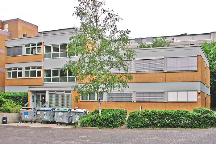
This home was found in a small concrete construction ideally situated right between the new buildings of Inorganic Chemistry and Biochemistry (formerly Institutes of Crystallography and Mineralogy) and within a stone's throw of the building of Organic Chemistry. The building was originally constructed to accommodate the electron microscopes of the Institute of Microbiology and Epizootics (Prof. Siegfried Grund) of the veterinary medicine department. None other than later Nobelist Ernst Ruska had planned two free-swinging bottom-up concrete pendulums on steel rods which are 8 meters deeply plugged into the ground to absorb vibrations from the nearby underground passing through Dahlem.
The Research Center of Electron Microscopy (FZEM) at the FU-Berlin was founded in 1999. A left-over Philips EM 400T (with STEM), a Philips CM12 and the new FEI Tecnai F20 build up the apparative equipment, when Dr. habil. Christoph Böttcher moved here with his group and became the first scientific head of the institute in July 1999.
... the Present, ...
But technical development has made significant progress, especially in recent years with new technologies such as direct electron detectors and volta phase plates. To keep up with this development, a 200 kV Talos Arctica with autoloader and VPP was funded from the DFG back in 2016 and took the place of the EM 400. Next a Talos L120C replaced the CM12 microscope and became, together with the Tecnai F20, our new working horse. In 2020, Dr. Böttcher retired and Dr. Kai Ludwig became Scientific head of the FZEM.
Besides, the building houses a Hitachi SU 8030 scanning electron microscope from the group of Prof. Dr. Eckart Rühl for inspections of mid-sized structures and a Bruker Multimode 8 nanoscope atomic force microscope from the group of Prof. Dr. Reiner Haag for investigations of nanostructured surfaces. While none of the older microscopes is present anymore, the two pendulums still do their job. One of them carries our Talos Arctica microscope, the other one balances the AFM.
Last but not least, we accommodate the concentrated knowledge of Natural Science by giving also the emeriti of the faculty a new home.
... and the Future.
Imaging supramolecular structures from synthetic amphiphiles is still one of our main concerns. However, from the side of structural biochemistry (esp. by the group of Prof. Markus Wahl) there was a growing need to use electron microscopy to elucidate the structures of biological macromolecules and macromolecular complexes in the nearly atomic resolution regime by means of the so-called single particle method (SPA). This requires a different instrumentation and so two 300 kV Titan Krios microscopes were acquired in a joint 91b application of Charité and FU-Berlin at the DFG in 2019; one specialised for cryogenic electron tomography (cryo-ET) in Berlin Buch and one for SPA data acquisition here in Dahlem. Already in the first year of routine operation, 3D structures of 100 to 1000 kD protein complexes have been achieved with resolutions up to 1.8 Å. Due to the rather huge dimensions of the Titan Krios, it could not find a place in our old building. Thus, it had to be temporarily housed in a large hall of the physics department.
With the completion of the new research building SupraFAB, the Titan Krios will move into it and will benefit there from the optimized, climate-stabilized and low-vibration structural conditions. As part of the Berlin Integrative Structural Biology (BIS), we will aim for a firm place in the growing field of 3D structural analysis of proteins and protein complexes.
Methodologies
- CTEM
- Cryo-TEM
- Cryo-Negative staining
- Cryo-Tomography
- SPA (Single particle data acquisition)
- 3D-Reconstruction (IMAGIC 5, CisTEM, IMod etc.)
Instrumentation
- Titan Krios with Falcon 3EC Direct Electron Detector
- Talos Arctica with CETA 4k CMOS camera and Falcon 3EC Direct Electron Detector
- Vitrobot Mark IV automated freeze plunger
- Talos L120C with CETA 4k CMOS camera
- Tecnai F20 FEG with Eagle 2k CCD camera
- Gatan Cryo Holder and -transfersystem (Model 626)
- Gatan Cryo High Tilt Tomography Holder and -transfersystem (Model 914)

