Instrumentation
In our investigations, we use the following instruments.
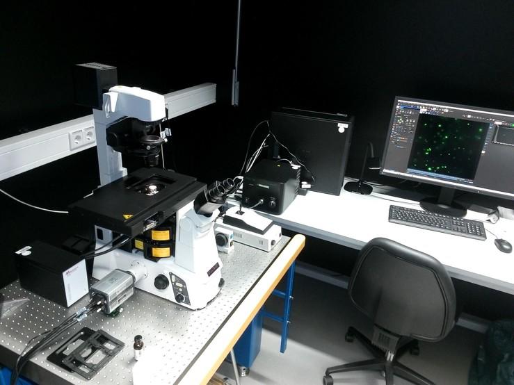
1.Nikon Ti-E Eclipse TIRF microscope
- Widefield transmission, fluorescence and total internal reflection fluorescence (TIRF)
- Fluorescence excitation by a gas discharge lamp, providing relatively homogenous intensity distributions over the entire field of view even in TIRF imaging
- Camera: Andor Zyla 4.2 Plus sCMOS camera connected via Camera Link (offering 100 fps at full resolution)
We have used this instrument to perform flourescence and TIRF imaging experiments off various samples ranging from the diffusion of fluorescently labeled nanoparticles (in bulk solution and at interfaces) to the motion of single, fluorescently labeled lipids in supported lipid bilayers.The combination of a powerful gas discharge lamp with a 3rd generation sCMOS camera achieves single-dye resolution for bright (high quantum yield) dyes, whereas dimmer samples (e.g., proteins labeld with a single GFP- or RFP-moiety) can be characterized using the laser TIRF system mentioned in the following. Our home-made microfluidic channels can be mounted on both TIRF systems.
The instrument is equipped with hardware-based focus stabilization, which keeps the glass-bulk interface (or any user-specified layer) in the focal plane during the measurement.Furthermore, the microscope contains a second level for sample excitation, which we currently use to add home-made interferometic scattering (iSCAT) imaging to this instrument.

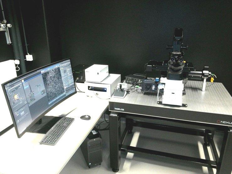
2. Nikon Ti2-E Eclipse Laser TIRF microscope
- Widefield transmission, fluorescence and total internal reflection fluorescence (TIRF)
- Fluorescence excitation by a gas discharge lamp
- TIRF excitation via a laserbox (405, 488, 561, 640 nm)
- Camera: Photometrics Prime BSI camera connected via USB 3 (offering 33 fps at full resolution)
- Hardware-based focus stabilization
This instrument is, from an applicability point of view, relatively similar to our Ti-E microscope. The combination of laser excitation with a 3rd generation back-illuminated sCMOS camera offers, however, single-dye resolution also for relatively dim dyes. Furtermore, a beam splitter (OptoSplit) can be inserted in the camera port, which allows to simultaneously record the fluorescence emission in two different fluorescence configurations. We will complement this instrument with an incubation chamber, which will add live cell imaging capabilities to this system.

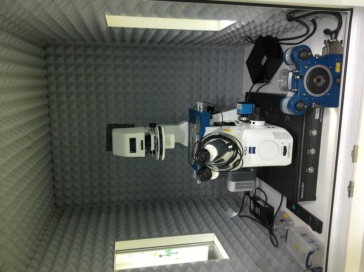
3. JPK NanoWizard 3 BioScience AFM + Zeiss AxioObserver.D1
- Atomic force microscope (AFM) mounted on an inverted fluorescence microscope
- Imaging modes (JPK): Contact and intermittend contact AFM modi (in air and liquid)
- Imaging modes (Zeiss, widefield): transmission, fluorescence
This instrument allows to characterize a sample area of interest using AFM and optical microscopy. It is equipped with the BioCell and the CellHesion module and is therefore applicable to a wide range of samples covering single molecules to living cells. In the past, we mainly used this instrument for force spectroscopic measurements of biomolecular interactions and for imaging of macromolecules and nanoparticles. Currently, we extend these investigations to a cellular context, e.g., by performing force spectroscopy on and imaging of cell membranes.

4. Interferometric scattering (iSCAT) microscopy
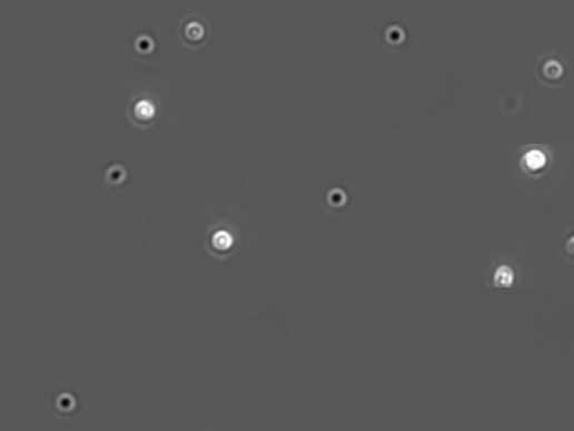
In order to improve the temporal resolution of our investigations, we recently started to focus on scattering microscopy. As scattering does not suffer from bleaching (in contrast to the dyes used in fluorescence microscopy), it can be operated at much higher illumination intensities and hence, at a much higher acquisition rate of the camera.
In our experiments we use a commercial solution (Refeyn OneMP), which images a sample area of approximately 10.8 x 10.8 µm² at a maximum acquisition rate of 1 kHz. In order to improve speed and size of the imaged area, we recently started to integrate a home-made iSCAT implementation into our Nikon Ti-E system.

