Stricker_02
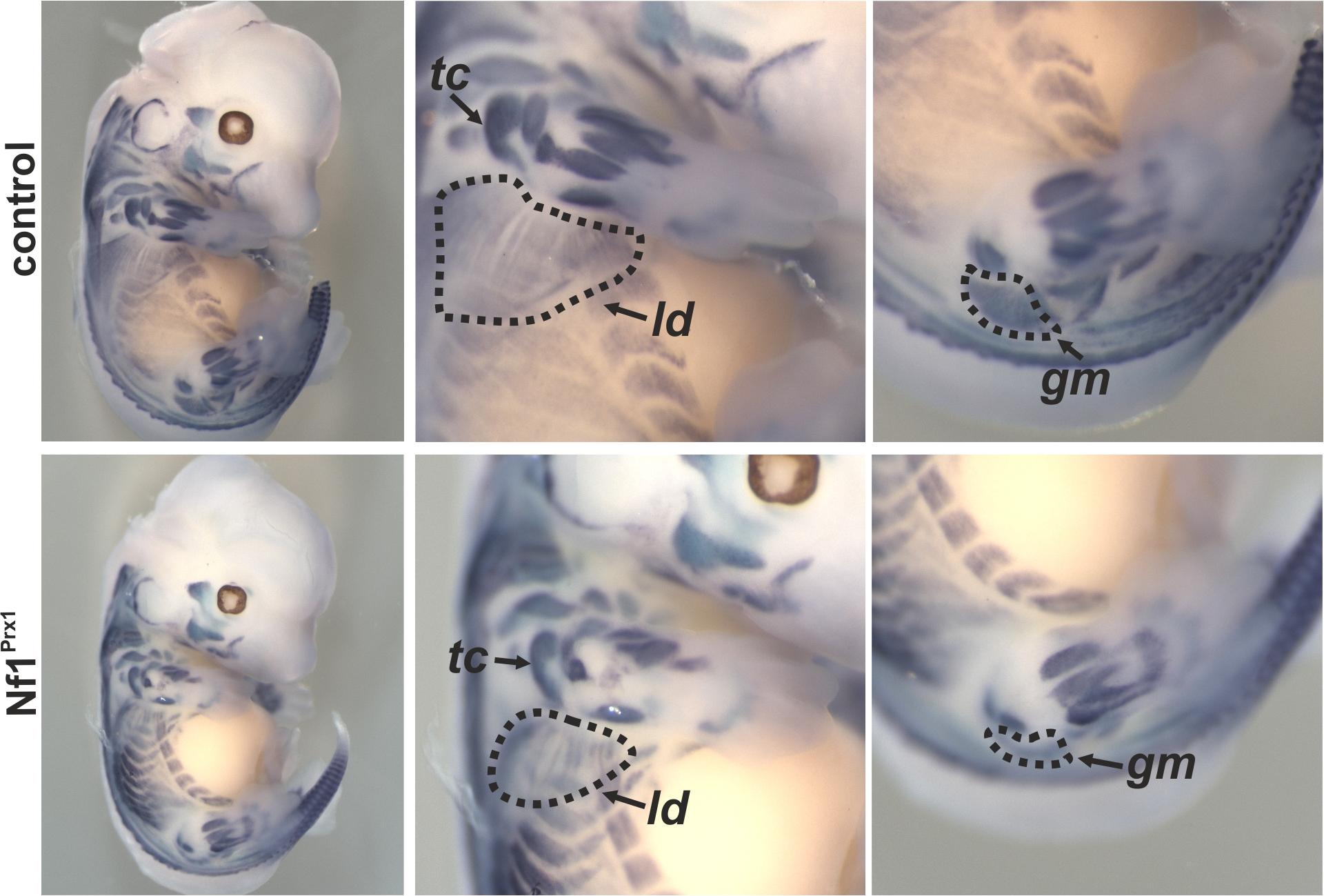
In-situ hybridisation for MyoD at embryonic day 13.5 demonstrating muscle patterning and size defects in the Nf1Prx1 mouse mutant. The magnifications show fore- and hindlimbs with reduction of muscle area visible e.g. for the latissimus dorsi muscle (ld), the triceps muscle (tc) or the gluteus maximus muscle (gm). Modified from Kossler et al. 2011.