
Gas-Phase Infrared Action Spectroscopy
Gas‑phase infrared (IR) action spectroscopy has emerged as a powerful tool for exploring the intrinsic structures of molecular ions with exceptional precision. By isolating nano-electrospray generated ions in the gas phase—free from the influences of solvents or surfaces—this technique allows us to probe the fundamental vibrational fingerprints of complex biomolecules and reactive intermediates. The obtained spectra enable the structural elucidation and assignment of the preferred conformations, in combination with ab initio calculations performed on the HPC facility at FUB-IT.
A major strength of gas‑phase IR action spectroscopy is its ability to characterize species that are difficult or impossible to study by condensed‑phase methods. This includes heterogeneous mixtures of isomers/conformers and extremely short‑lived reactive intermediates. In our research group, we apply this technique to structurally challenging molecular classes such as glycans, metabolites, peptides, nucleotides, and glycosyl cations. Glycans, for example, often differ only subtly in their branching patterns or stereochemistry, yet these small variations can have profound biological consequences. Gas‑phase IR spectra provide a direct structural readout that helps disentangle these complexities. Similarly, metabolites and peptides—often present as mixtures of isomers or conformers—can be distinguished through their unique vibrational signatures. Perhaps most strikingly, gas‑phase IR action spectroscopy enables the characterization of glycosyl cations, reactive intermediates central to glycosylation chemistry, which cannot be isolated or observed in solution.
In cooperation with the group of Gert von Helden from the Fritz Haber Institute (FHI) of the Max Planck Society, our group employs two forms of cryogenic gas‑phase infrared action spectroscopy on custom-built instruments, each designed to achieve high spectral resolution and sensitivity by cooling ions to extremely low temperatures:
1. Messenger‑tagging spectroscopy of mass‑selected and mobility‑separated ions at 45 K.
In this approach, mass‑selected and mobility‑separated ions are cooled in a cryogenic ion trap at 45 K and weakly bound “messenger” molecules such as N₂ or rare‑gas atoms are attached. Absorption of an IR photon causes the messenger to detach, and this loss is monitored as the spectroscopic signal. When combined with ion mobility separation, this method allows us to isolate and characterize individual conformers or isomers within complex mixtures. Experiments are performed either on the Cold-Photo-Synapt (a modified Synapt G2-S traveling-wave ion mobility-mass spectrometer) located at the SupraFAB Research Building of the FU Berlin, or on the iMob (a home-built instrument consisting of a drift tube) located at the FHI. Tunable infrared radiation for these experiments are generated using benchtop optical parametric oscillator/optical parametric amplifier (OPO/OPA) systems.

Figure: Schematic representation of the Cold-Photo-Synapt instrument.
2. Helium nanodroplet spectroscopy at 0.4 K.
In this approach, mass‑selected ions are gently captured inside superfluid helium nanodroplets, which cool them to an ultralow temperature of approximately 0.4 K. At such temperatures, thermal broadening is minimized, and the resulting IR spectra exhibit sharp, well‑resolved features that reveal structural details. The helium environment is remarkably gentle. It stabilizes the ions without significantly perturbing their intrinsic structures. Experiments are performed on a home-built instrument located at the FHI. Tunable infrared radiation for these experiments is provided by the FHI free electron laser (FHI-FEL).
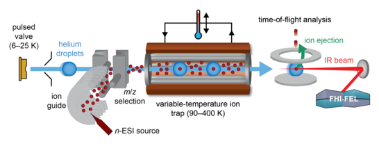
Figure: Schematic representation of the HeDrop instrument.
Related Publications
- Cryogenic Gas-Phase IR Spectroscopy on a Commercial Ion Mobility-Mass Spectrometry Platform
Szekeres, G.P.; Jordan, J.S.; Riedel, J.; Horlebein, J.; Prabhu, G.R.D.; Götze, M.; Daly, S.; Warnke, S. & Pagel, K. Anal. Chem. 2026 - Cryogenic Gas-Phase Infrared Ion Spectroscopy of Ultraviolet-Induced Nucleotide Photoproducts
Prabhu, G.R.D.; Götze, M.; Greis, K.; Torres-Boy, A.Y.; Safferthal, M.; Strzelecka, D.; Kirschbaum, C.; Deshpande, N.D.; Geue, N.; Meijer, G.; von Helden, G. & Pagel, K. Anal. Chem. 2025 - Distinguishing Isomeric Caffeine Metabolites through Protomers and Tautomers Using Cryogenic Gas-Phase Infrared Spectroscopy
Geue, N.; Prabhu, G.R.D.; Renzi, E.; Walton-Doyle, C.; Meijer, G.; von Helden G. & Pagel, K. Anal. Chem. 2025 - The Impact of Side-Chain Fluorination on Proton-Bound Phenylalanine Dimers: A Cryogenic Infrared Spectroscopic Study
Safferthal, M.; Greis, K.; Change, R.; Chang, C.W.; Hoffmann, W.; Meijer, G.; von Helden, G. & Pagel, K. Phys. Chem. Chem. Phys. 2024 - Predicting Structural Motifs of Glycosaminoglycans Using Cryogenic Infrared Spectroscopy and Random Forest
Riedel, J.; Lettow, M.; Grabarics, M.; Götze, M.; Miller, R.L.; Boons, G.J.; Meijer, G.; von Helden, G.; Szekeres, G.P. & Pagel, K. J. Am. Chem. Soc. 2023 - Studying the Key Intermediate of RNA Autohydrolysis by Cryogenic Gas-Phase Infrared Spectroscopy
Greis, K.; Kirschbaum, C.; Taccone, M.I.; Götze, M.; Gewinner, S.; Schöllkopf, W.; Meijer, G.; von Helden, G. & Pagel, K. Angew. Chem. Int. Ed. 2022 - Unravelling the Structural Complexity of Glycolipids with Cryogenic Infrared Spectroscopy
Kirschbaum, C.; Greis, K.; Mucha, E.; Kain, L.; Deng, S.; Zappe, A.; Gewinner, S.; Schöllkoph, W.; von Helden, G.; Meijer, G.; Savage, P.B.; Marianski, M.; Teyton, L. & Pagel, K. Nat. Commun. 2021 - Direct Experimental Characterisation of the Ferrier Glycosyl Cation in the Gas Phase
Greis, K.; Kirschbaum, C.; Leichnitz, S.; Gewinner, S.; Schöllkopf, W.; von Helden, G.; Meijer, G.; Seeberger, P.H. & Pagel, K. Org. Lett. 2020 - Unravelling the Structure of Glycosyl Cations via Cold-Ion Infrared Spectroscopy
Mucha, E.; Marianski, M.; Xu, F.F.; Thomas, D.A.; Meijer, G.; von Helden, G.; Seeberger, P.H. & Pagel, K. Nat. Commun. 2018 -
Glycan Fingerprinting via Cold-Ion Infrared Spectroscopy
Mucha, E.; Gonzàles Flórez, A.I.; Marianski, M.; Thomas, D.A.; Hoffmann, W.; Struwe, W.B.; Hahm, H.S.; Gewinner, S.; Schöllkopf, W.; Seeberger, P.H.; von Helden, G. & Pagel, K. Angew. Chem. Int. Ed. 2017


